SKYCAM Confocal Microscopy
SKYCAM Confocal Microscopy
Description
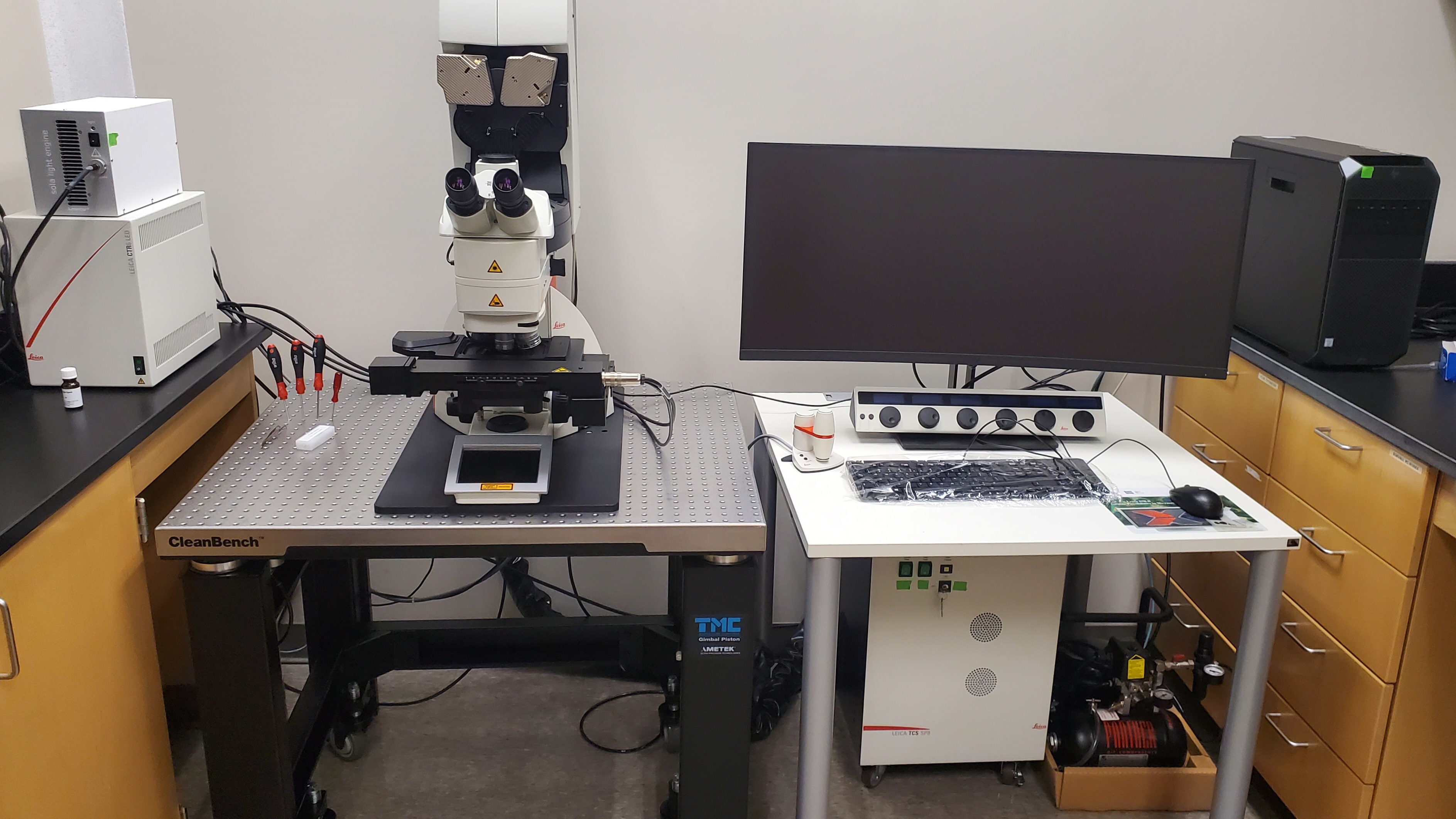
- Leica TCS SP8 Laser Scanning Confocal Microscope with four laser lines and associated optics. The microscope was acquired through a Major Research Instrumentation Grant funded by the National Science Foundation in 2019 with Dr. Srivastava as the PI.
Capabilities of Microscope
- The microscope can perform routine scanning, Z-Stacking, Unmixing of spectral overlap,
post-acquisition image processing.
- Available Laser Lines: 405 nm, 488 nm, 552 nm, 638 nm.
- Available Objectives: 10X, 20X Immersion, 40X Water Immersion, 63X Oil. All objectives Plan Apo.
Technical Description
- DM6 CS 5 pos fluo
- Optical Outfit + ICT
- SolaSEII 365nm
- Transmitted Light Brightfield Detector
- Scanning stage DM6 CS
- Scan optics module HIVIS with rotation
- FOV scanner SP8
- SP8 LIAchroics Compact RGB
- Four Internal Detector Channels pos. 1, 2, 3, 4
- Internal Detector Channel PMT 1
- Internal Detector Channel HyD 2
- Internal Detector Channel PMT 3
- Internal Detector Channel HyD 4
- Laser 405 nm DMOD Compact
- Laser 488 nm, 552 nm, 638 nm
- Confocal Super-Resolution LAS X LIGHTNING
- Premium CUDA Workstation, Monitor
- Computer table small, 80x80 cm
- Confocal module with patented filter-free spectral Leica SP detector for up to five individually regulatable channels.
- Highly efficient spectral separation by unique prism design.
- Equal brightness between channels due to W-shaped slit design.
- Leica LIAchroics are low incident angle dichroic beam splitters custom designed by Leica Microsystems inhouse.
- The LIAchroic approach allows steeper cutoff and higher transmission than other dichroic beam splitter designs resulting in high contrast images.
- Scan optics with HIVIS coating for maximal transmission from 400-800 nm.
- The LIGHTNING image information extraction package serves for maximum extraction of image details and for confocal maximum resolution, thus expanding the imaging portfolio both in the classical range and beyond the diffraction limit. Even with an extremely low signal-to-noise ratio, LIGHTNING enables structures to be extracted from data sets that would otherwise not be visible or accessible. On the other hand, structures up to 120nm in several colors can be resolved simultaneously and at high recording speeds.
Service Fees
- WKU Users: $25/hour
- External Users: $40/hour
Please contact Dr. Ajay Srivastava (ajay.srivastava@wku.edu) for more details.
Confocal Contact
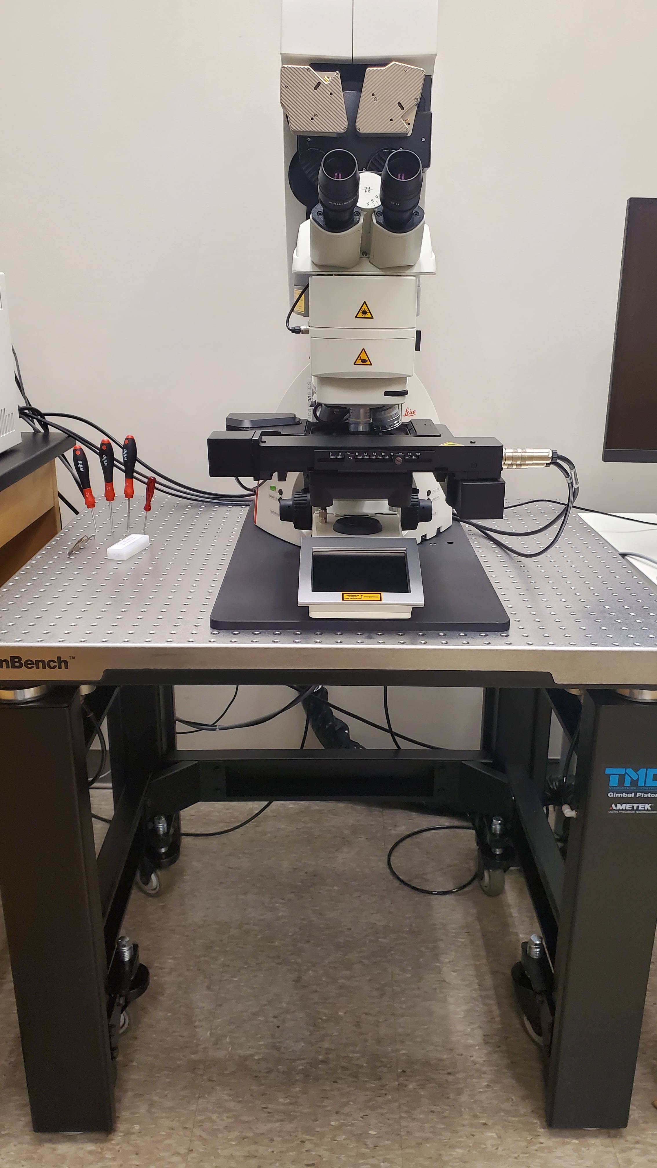
Confocal Microscope Front View
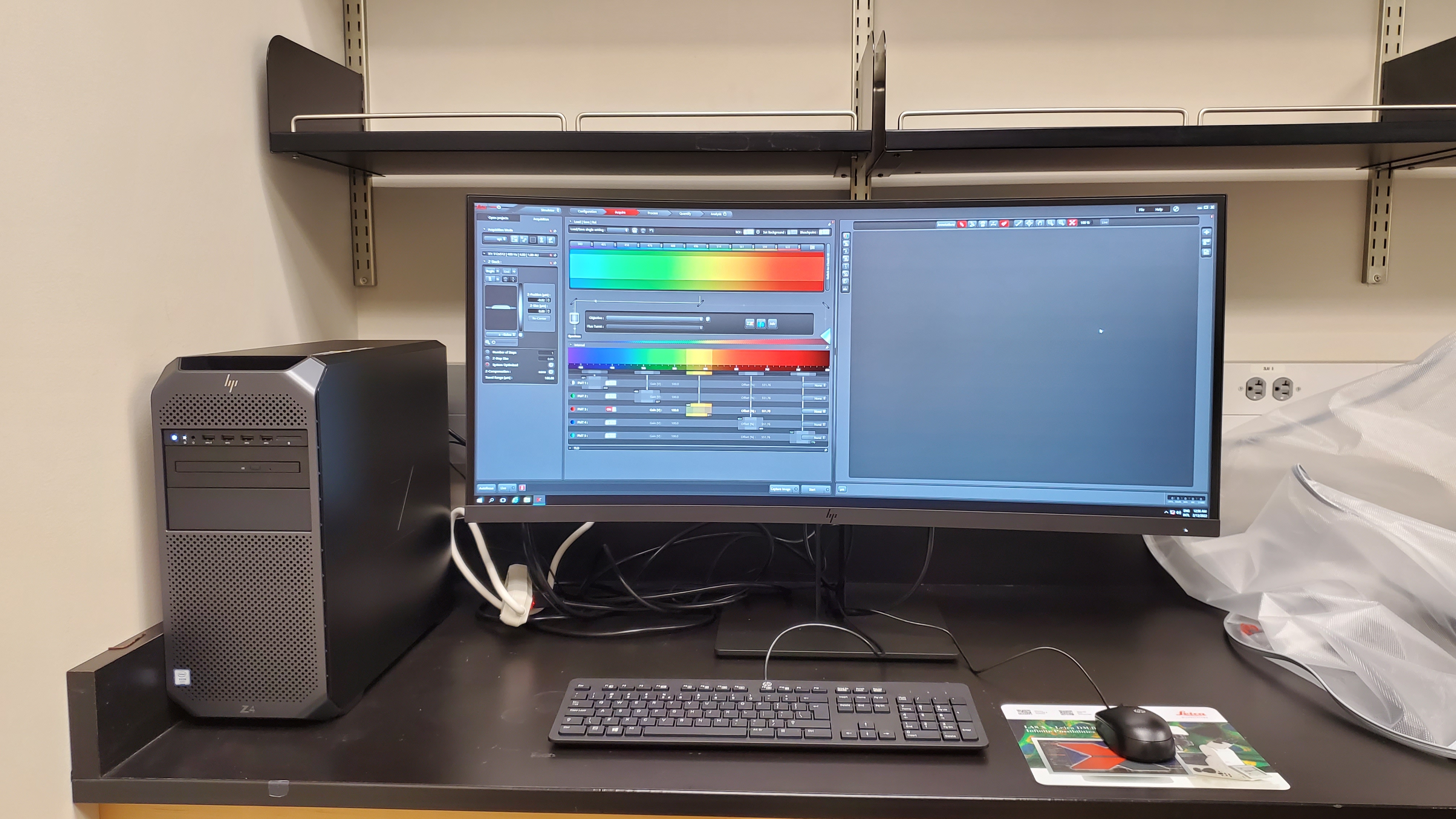
Confocal Microscope Computer
Confocal Microscope & Computer
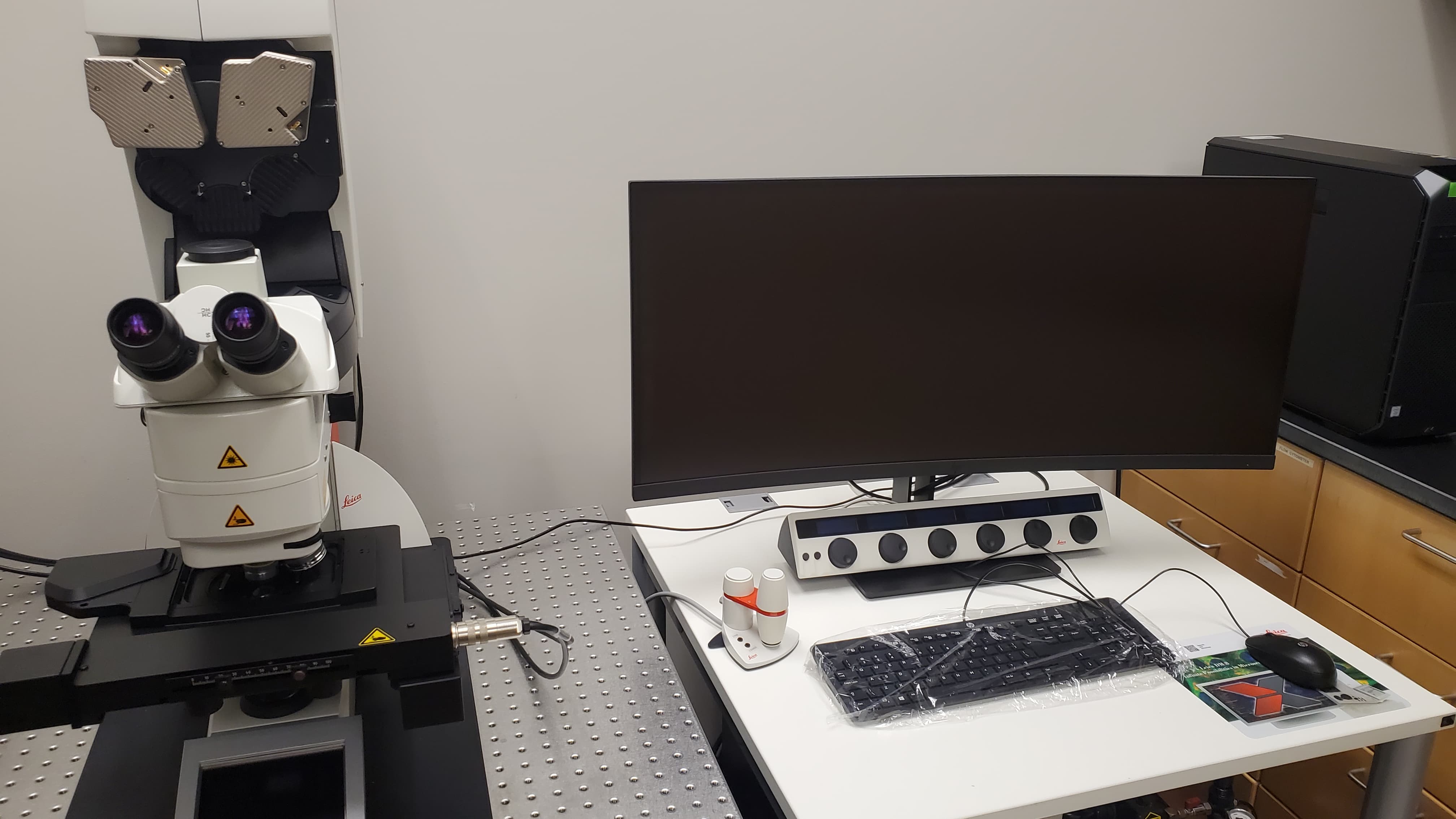
Confocal Microscope & Computer (Front View)
SKYCAM Microscopes
Agilent 5500 Atomic Force Microscope for surface imaging at the nanometer scale & Nanosurf FlexAFM Microscope for surface imaging below the nanometer scale.
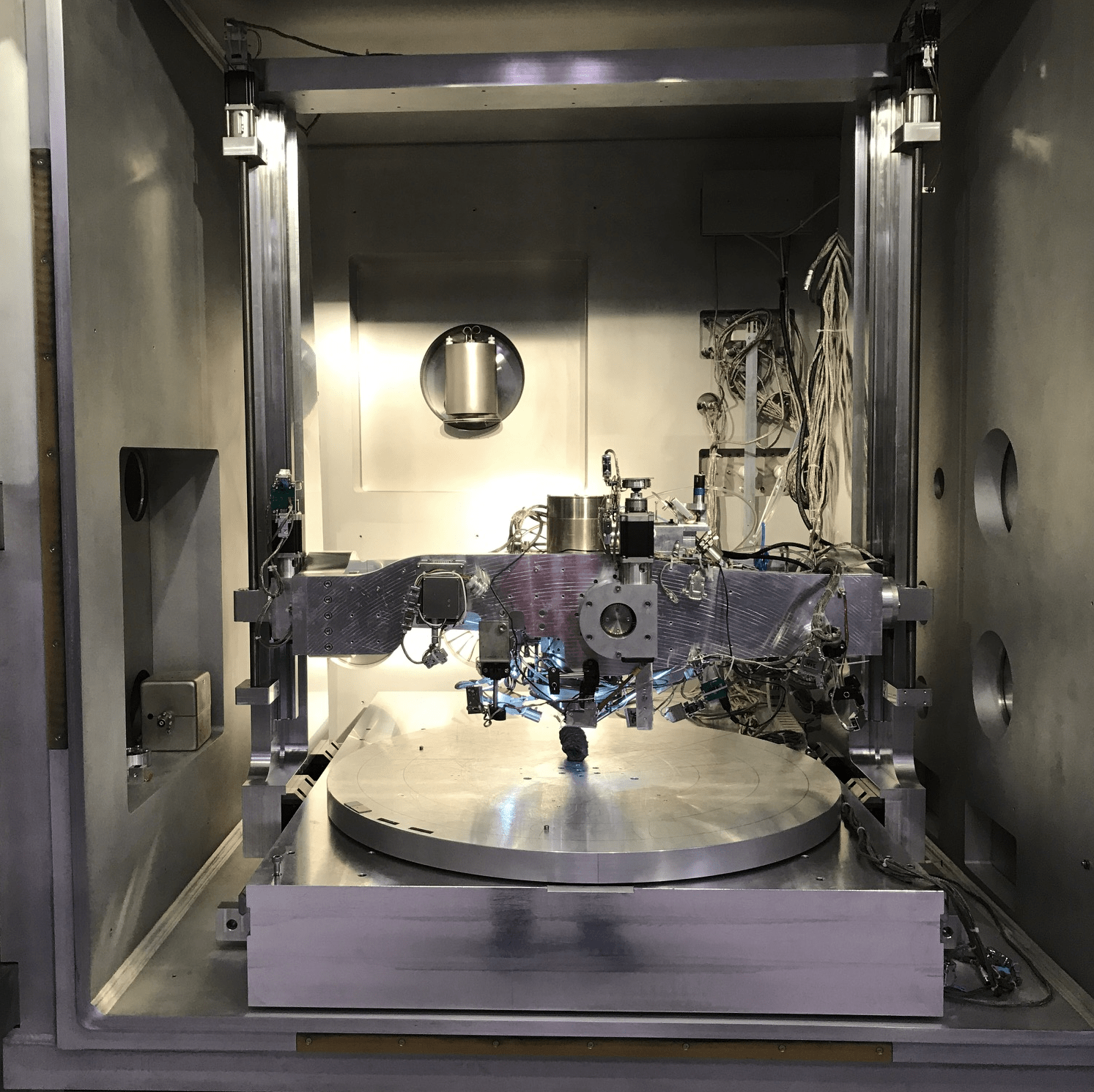
Unit can handle complete parts up to 1.5 meters Square and weighing up to 600 lbs. of weight EDX, EHT and Backscatter Detectors are on board with other types available EBDX (crystal structure) ECT.
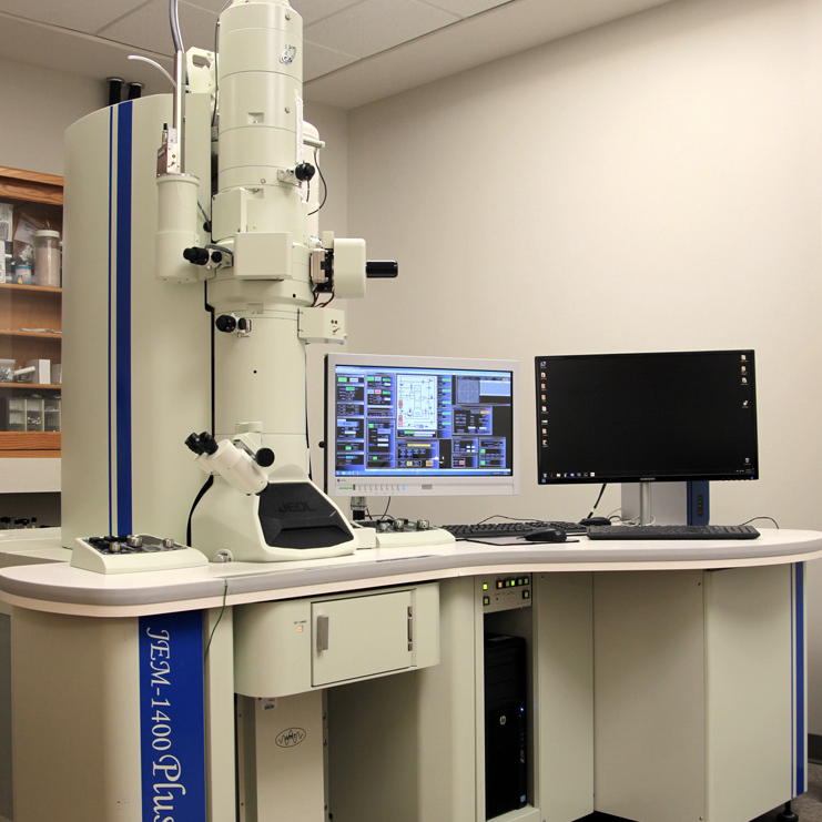
JEOL 1400PLUS 120kV Transmission electron microscope with an IXRF energy dispersive x-ray spectrometer and cryo-fin & JEOL 6510LV 30kV Scanning electron microscope with an IXRF energy dispersive X-ray spectrometer supporting both secondary electron and backscattered electron imaging.
Some of the links on this page may require additional software to view.