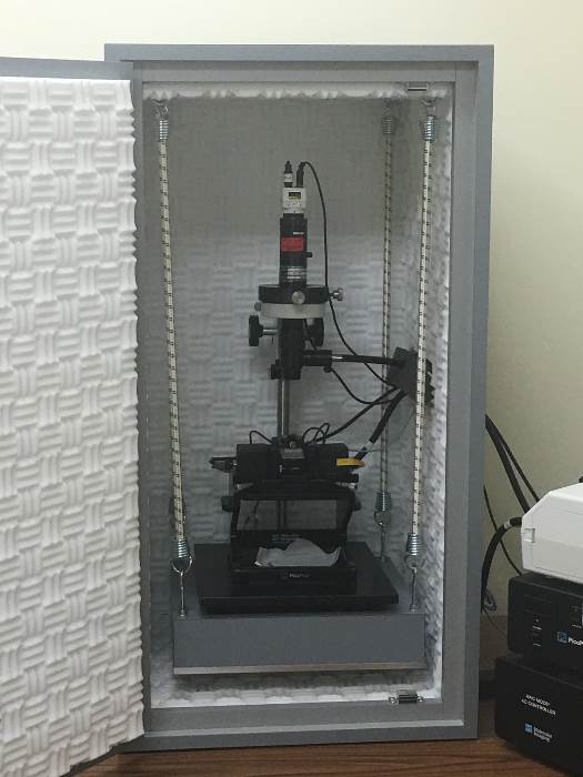
Atomic Force Microscopy (AFM)
|
Atomic Force Microscopy (AFM) is a technique for imaging the surface topography of solid materials at near atomic resolution. Piezoelectric crystals are used to direct tiny but accurate movements of a cantilevered sharp probe in a scan across the surface of the material sample. Forces between atoms on the surface and atoms in the tip of the probe are detected by measuring the deflection of the probe perpendicular to the surface. The resulting AFM image is a display of the height (z) of the surface as a function of position (x,y) on the surface, and is commonly represented as a pseudocolor plot. Typical AFM applications include surface characterization of a wide range of materials including metals and alloys, semiconductors, polymers and thin-film samples. Unlike an electron microscope, the sample does not need to be in a vacuum, allowing AFM images to be obtained from cells and many other forms of biological materials which would not survive a vacuum environment. Instrumentation: Agilent 5500 AFM Specifications: X,Y Scan range: 90 mm ´ 90 mm X,Y Noise level: 0.05 nm ´ 0.05 nm Z Scan range: 8 mm Z Noise level: 0.02 nm
Agilent 5500 AFM |
Some of the links on this page may require additional software to view.


